What is a Dental Bone Graft?
A dental bone graft is a procedure that adds bone or a bone-like material to areas of the jaw where bone is thin or missing. The graft provides a scaffold so your own bone cells can grow and strengthen the site. Dentists use it to preserve bone after an extraction or to prepare a stable foundation for future treatment, such as implants.
You lost a molar years ago, and the ridge has thinned. When a tooth is missing, the jawbone can shrink over time. Gum disease, infection, or injury can also reduce bone volume. Because of this, a graft helps restore support for chewing and for planned restorations.
- Preserve the socket after a tooth extraction
- Increase ridge width or height before implant placement
- Lift the sinus floor for upper back implants (sinus augmentation)
- Repair defects from gum disease around teeth
- Rebuild areas after cyst removal or trauma
Grafts can come from your own bone (autograft) or from donated human, animal, or synthetic sources that are carefully processed for safety. The material holds space and guides new bone growth. Healing usually takes several months, depending on the site and the material used. Both horizontal and vertical ridge augmentation approaches can rebuild atrophic ridges when properly selected [1]. Sinus floor elevation, used in the upper back jaw, has shown favorable long-term implant outcomes in systematic reviews [2]. Emerging options, such as autologous dentin grafts, have reported promising regenerative results in analyses [3].
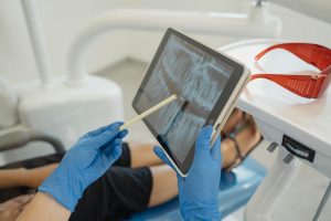
Your dentist will examine the area, review your medical history, and may use 3D imaging to plan the graft. If an implant is part of your plan, see our overview of the implant healing timeline to understand next steps. For visit details and availability, check our current hours. A stable foundation supports lasting dental work.
Why is Dental Bone Grafting Needed?
Bone grafting is needed when the jaw has lost volume or density and can no longer support teeth, implants, or gums well. It restores the foundation so chewing forces can be handled safely and so planned treatments, like implants or certain bridges, have stable support. A dental bone graft can also help keep the shape of your jaw after a tooth is removed.
After a tooth is lost, the bone that once held it begins to shrink because it no longer gets regular stimulation from chewing. Over time, this can leave the ridge too thin or soft for an implant or even for a well-fitting denture. Grafting replaces missing structure and signals your body to build new bone in that space. Because of this, it helps protect nearby teeth from drifting and reduces food trapping in deep defects.
In certain cases, grafting is part of periodontal care. Deep bone defects from gum disease weaken tooth support; filling those areas with graft material encourages regrowth and can improve tooth stability. For the upper back jaw, the sinus can slowly expand into the empty space after extractions, reducing bone height. A sinus lift adds height back so an implant can be placed where it is needed.
Timing also matters. Placing a graft right after an extraction can preserve the socket’s shape, making later treatment more predictable. If bone loss has already occurred, staged grafting may be recommended first, with implants or other restorations planned after healing. You plan an implant where bone shrank after years without a tooth. In that scenario, rebuilding the site first can make the final result stronger and more comfortable to use. To see how replacement choices fit into a broader plan, you can also compare tooth replacement options.
Understanding why grafting is advised helps you weigh steps, healing time, and long-term function with your dentist. Joint care between dental and medical teams helps sustain long-term wellness.
Understanding Ridge Preservation Techniques
Ridge preservation techniques are steps taken at or soon after a tooth extraction to help maintain the jaw’s shape and volume. They often pair careful, minimally traumatic tooth removal with placing a graft in the socket and sealing the site so it heals with more predictable contours. The goal is to keep bone where you may later want an implant or another restoration.
After an extraction, the body remodels the socket, and the outer bone wall can shrink more than the inner wall. Ridge preservation works by holding space with graft particles, protecting the blood clot, and limiting collapse while your bone fills in. Covering the socket with a membrane or a collagen plug can support better contour and reduce soft tissue growing into the space, which may improve dimensional stability compared with leaving it open [4].
Several materials can be used as the scaffold, including processed donor, animal-derived, or synthetic options, paired with resorbable membranes when indicated. Some dentists also add platelet concentrates, such as PRF or CGF, mixed with the scaffold to deliver growth factors. Systematic reviews report that incorporating these biologics with guided bone regeneration can enhance early bone fill and graft stability in appropriate cases [5].
You had a cracked premolar removed this morning. In many cases, a simple socket graft with a collagen seal is enough to preserve width for a future implant. When the bone around the roots is thin, the site may be covered and sutured for added protection. If conditions are ideal, an immediate implant can be placed with gap grafting; otherwise, the implant is planned after healing. A dental bone graft may be part of either approach. To see how healing unfolds week by week, review our extraction healing timeline. Prompt planning supports stable healing and reliable restoration.
The Sinus Lift Procedure Explained
A sinus lift, or sinus augmentation, adds bone to the upper back jaw so an implant can be placed safely. The sinus membrane is gently raised, and graft material is packed into the space to increase bone height. This procedure is a type of dental bone graft designed for areas beneath the maxillary sinus.
You need an upper molar implant but the bone is too short. This often happens after extractions when the sinus slowly expands into the empty space. A sinus lift rebuilds that height so the implant can anchor into strong, healthy bone.
There are two main approaches. In a lateral window lift, a small opening is made on the side of the ridge to access and elevate the sinus membrane, then graft is placed and the site is closed. In a crestal, or osteotome, approach, access is made from the top of the ridge through a small channel, and the membrane is carefully lifted from inside the implant site. Your dentist selects the method based on remaining bone, tooth position, and imaging.
What you might experience is usually mild pressure, swelling, or bruising for a few days. Many patients are advised to avoid pressure changes in the nose and sinuses while the area stabilizes. The graft then remodels over several months as your body forms new bone. Depending on stability and bone quality, the implant may be placed at the same visit or after healing.
As with any surgery, there are risks, such as a small tear in the sinus membrane, infection, or graft movement. Understanding these possibilities helps you plan timing, travel, and follow-up. If you are comparing your overall implant plan, you may want to review implant risks and considerations. Clear goals, good imaging, and communication support a smooth path to restoration.
Careful planning here can make your future implant more predictable and comfortable.
Meet with our team to plan the right implant option and timeline.
A confident smile starts with a single consultation.
What is Guided Bone Regeneration (GBR) Graft?
Guided bone regeneration is a dental bone graft technique that uses a barrier membrane to protect and shape a healing space where new bone can form. The dentist places graft material and then covers it with a membrane that keeps fast-growing soft tissue out while slower-growing bone cells fill in. GBR is used to rebuild jaw width or height around natural teeth or implants.
The membrane creates a secluded, stable chamber, helping the blood clot and graft stay in place as your body lays down new bone. Membranes may be resorbable or non-resorbable, and they are secured with tacks or sutures to prevent movement. Surgeons select grafts such as processed donor, animal-derived, or synthetic particles for space making; in some cases, tenting screws or small plates support the shape. Healing and remodeling often take several months, adjusted to defect size and biology. Systematic reviews describe favorable augmentation outcomes that support predictable implant placement in properly selected cases [6]. Long-term evaluations report that bovine xenograft particles can integrate and help maintain volume in oral surgery sites [7].
Planning determines whether GBR is performed at the same time as implant placement or in stages. If the implant has strong initial stability but a thin wall, a small dehiscence can be covered with graft and membrane. When bone is too narrow or short, staged GBR first builds structure, then the implant follows after healing. Your dentist finds a thin outer bone wall during implant placement. Some teams add platelet-rich fibrin to the graft complex to support early bone fill, as summarized in recent meta-analyses [8]. In select peri-implantitis defects, regenerative approaches with membranes and grafts can achieve radiographic bone gain when conditions are favorable [9].
For patients, GBR aims to restore strength where bone is missing so chewing forces transfer safely to the jaw. Understanding the steps, materials, and healing timeline helps set clear expectations and improve comfort during recovery. If implants are part of your plan, you can explore how implant healing works to see how these phases fit together. Thoughtful GBR planning supports long-term function and comfort.
Bone Graft Cost Considerations
Bone graft cost depends on the size of the defect, the type of graft, and how complex the surgery is. Fees also reflect the number of sites treated, the need for membranes or fixation, and whether the procedure is staged or combined with an implant. Because every case is unique, your dentist will outline a plan and the associated range before treatment. A dental bone graft is priced to match the materials, time, and follow-up care required.
You need a sinus lift and a ridge graft in one area. Larger or multi-wall defects take more time and material than a simple socket graft, which raises cost. Autografts can add surgical time because a second site is prepared, while processed human, animal, or synthetic options avoid that step but still vary in procurement and handling needs. Barrier membranes, tenting screws, or pins add to material use and chair time. Sedation and advanced imaging, such as CBCT and surgical guides, are separate line items in many treatment plans.
Timing influences the total as well. When an implant is placed at the same visit and conditions are ideal, some visits can be combined. If the site is thin or infected, staged grafting first is safer, though it adds appointments. Health factors like smoking, uncontrolled diabetes, or certain medications can slow healing, which may increase follow-up care. Related procedures, such as extraction, sinus lift, or periodontal therapy, are typically billed separately and included in the overall plan.
Coverage varies. Some dental plans limit benefits for grafting tied to implants, while medical plans may consider grafts after trauma, cyst removal, or congenital defects when documentation is provided. Pre-authorization helps clarify your out-of-pocket portion before you start. For broader budgeting around implant treatment, you can compare implant cost factors and financing approaches. Clear planning keeps treatment efficient and helps you avoid surprises.
Grafting After Tooth Extraction
Grafting after a tooth extraction means placing a small amount of graft material into the empty socket to help preserve bone. The material acts as a scaffold while your body replaces it with new bone. This approach can keep the ridge closer to its original shape for future treatment. A dental bone graft at the time of removal often simplifies later planning.
Without a graft, the socket naturally shrinks, most noticeably on the outer wall of the jaw. Socket grafting limits that collapse by holding space and protecting the healing clot, so more bone volume remains for implants or other restorations. Systematic reviews show that alveolar ridge preservation maintains more bone compared with letting the site heal on its own [10]. In the back upper jaw, preserving height and width can be especially helpful for later implant placement [11].
Your lower molar cracks and must be removed today. After the tooth comes out, the dentist gently cleans the socket, places graft particles, and seals the site with a collagen plug or membrane, then sutures. The area heals over several weeks while the graft integrates and remodels. Many patients return for imaging and treatment planning once the site is stable. The exact timing depends on biology, site size, and your overall plan.
Not every socket needs grafting, and decisions vary in wide molar sites, where evidence suggests case-by-case assessment rather than a universal rule [12]. If a wall is missing or infection is present, a staged regenerative approach may be safer. When immediate implants are placed, a small gap graft can support contour around the fixture. Good home care, avoiding smoking, and keeping follow-up visits support predictable healing; for day-by-day expectations, see our recovery timeline. Thoughtful timing preserves options for the restoration you want later.
Recovery and Aftercare for Bone Grafts
After a dental bone graft, expect mild swelling, bruising, and soreness for a few days. Keeping the site stable and clean helps the graft succeed. Gums usually feel better within 1 to 2 weeks. New bone forms slowly and can take several months to mature.
For the first day, rest with your head raised and use cold packs 10 minutes on, 10 minutes off. Bite gently on gauze to control oozing, and avoid spitting, swishing hard, or using a straw. Choose soft, cool foods while the area is tender. A common real-world example: yogurt and eggs for breakfast work well. After the first 24 hours, you can start gentle warm saltwater rinses, unless told otherwise.
Brush and floss the other teeth as usual, but do not scrub the graft site. Do not pull at your lip or cheek to “look” at it. If a membrane or white film shows a little, leave it alone. Take pain medicine only as directed, and start any antibiotic or antimicrobial rinse if it was prescribed. Avoid smoking and alcohol, which slow healing.
If your graft was near the sinus, skip nose blowing and try to sneeze with your mouth open for about a week. Small sand-like particles in your saliva can happen early on and are usually harmless. Stitches may dissolve on their own or be removed in 1 to 2 weeks, based on the material. Call us if bleeding soaks gauze for more than a few hours, swelling or pain increases after day three, you develop fever, or you notice pus or a foul taste.
Follow-up visits let your dentist check healing and plan the next steps. Soft tissue closes first, then the graft remodels into stronger bone over 3 to 6 months, depending on location and biology. For comfort expectations, you can review what normal post-extraction pain feels like. Coordinated care often leads to smoother healing and better function.
Potential Risks of Dental Bone Grafting
Any surgery has risks, and bone grafting is no different. Most people experience temporary swelling, bruising, and soreness. Less commonly, problems include infection, gum opening over the graft, or the graft not fully joining with your bone. Rare issues depend on the site, such as sinus symptoms after a sinus lift or nerve irritation in the lower jaw.
Infections can start if bacteria enter the site, especially when the gum edges separate or a barrier membrane becomes exposed. Movement at the graft, smoking, and uncontrolled diabetes increase the chance of delayed healing. Your dentist reduces these risks with sterile technique, stable flap closure, and instructions that limit pressure and motion in the area. Good home care and follow-up visits also matter. Most early concerns respond to cleaning, local care, or medicine when caught quickly.
Sinus augmentation has its own considerations. A small tear in the sinus membrane can occur during elevation, and some patients develop short-term sinus pressure or congestion. You sneeze after a sinus lift and feel pressure on one side. Surgeons aim to prevent this with careful technique, and they advise you to avoid nose blowing and heavy straining while the area heals. Controlled trials in a systematic review outline these potential complications and support careful case selection to limit them [13].
Block grafts and larger reconstructions carry added surgical steps and, at times, higher chances of wound opening, graft exposure, or partial loss. Donor site soreness is possible if your own bone is used. A systematic review of bone block use in practice highlights these risks and the importance of tension-free closure and immobilization for success [14]. Allergic reactions to processed graft materials are rare, since they are treated to remove cells and proteins. Nerve changes are uncommon, but your team plans around nerve locations to keep them safe.
Call your dentist if bleeding is heavy, pain or swelling worsens after a few days, you see pus, or you develop fever. Understanding risks helps you act early and protect the graft while it heals. Report concerns early so small issues do not become setbacks.
Long-Term Success Rates of Bone Grafts
Most dental bone grafts support stable, long-lasting results when planned and maintained well. In implant dentistry, grafted sites can achieve high implant survival over many years, though outcomes vary by defect size, graft type, and timing [15]. For sinus lifts, systematic reviews report favorable long-term performance with careful case selection and technique [2].
Why do grafts succeed long term? They create a stable, well-vascularized space that your body can remodel into strong bone. Primary wound closure, minimal micromotion, and healthy gums protect the site as it matures. Patient factors matter too. Non-smoking status, good plaque control, and stable medical conditions support durable bone and healthier implants over time.
The starting condition of the jaw guides expectations. Small socket or ridge defects often remodel predictably. Larger reconstructions can also work well, yet they demand meticulous planning and longer healing. For example, after microvascular fibula flap reconstruction of the jaws, many patients achieve functional dental rehabilitation, but prior radiation, soft tissue quality, and prosthetic design influence outcomes [16]. These variables help explain why one person may need staged grafting while another can combine steps safely.
Sinus augmentation is a common long-term success story. When bone height is limited in the upper back jaw, elevating the sinus floor and placing a graft can rebuild sufficient height for a stable implant platform years later [2]. Comparable principles guide guided bone regeneration, ridge augmentation, and other approaches, all aiming for space maintenance, blood supply, and tension-free closure. A dental bone graft is a means to a functional, maintainable foundation, not an end by itself.
Real-world snapshot: a thin upper molar ridge is grafted today, and years later the implant still feels solid. Your long-term result depends on thoughtful diagnosis, steady home care, and regular follow-up. Ask how your specific site, materials, and timing shape the expected timeline and durability. Consistent hygiene and checkups help preserve your investment for the long run.
Frequently Asked Questions
Here are quick answers to common questions people have about Dental Bone Grafts Explained in Glendale, AZ.
- What types of bone graft materials are used in dental procedures?
Dental bone graft materials can come from various sources. Commonly used are:
- Autografts: Bone taken from your own body.
- Allografts: Processed human bone from a donor.
- Xenografts: Bone from animals, typically bovine.
- Synthetic options: Man-made materials like calcium phosphates.
Each type has different benefits and risks. Your dentist will choose the best material based on your need and health condition.
- Is dental bone grafting painful?
Pain from a dental bone graft is usually mild to moderate. You’ll likely experience soreness, swelling, and some bruising, which are common after surgical procedures. Your dentist can prescribe pain medication and offer tips to manage discomfort. Cold packs for swelling and rest can speed recovery. Most patients report that discomfort is manageable and decreases over a few days. Following post-operative care instructions can make the recovery process smoother.
- How long does it take to heal after a dental bone graft?
Healing after a dental bone graft typically takes several months. Initial gum healing often happens within 1 to 2 weeks, but new bone formation can take 3 to 6 months or longer. The duration depends on the graft site, material used, and your overall health. It’s important to follow your dentist’s instructions and attend follow-up visits to ensure proper healing and monitor progress.
- Can a dental bone graft fail?
Yes, dental bone grafts can fail, although this is uncommon. Potential failures could be due to infection, movement at the graft site, smoking, or certain underlying health conditions like diabetes. Careful surgical technique and following aftercare instructions reduce these risks. Your dentist will closely monitor the healing process and provide guidance to promote graft success. Early detection and treatment of any issues can help avoid failure.
- Is smoking a risk factor for bone grafting success?
Yes, smoking is a significant risk factor that can affect the success of a dental bone graft. It reduces blood flow and slows healing, increasing the chance of complications like infection or poor graft integration. Quitting smoking before and after a bone graft procedure greatly improves the likelihood of a successful outcome. Your dentist may provide resources to help manage smoking cessation as part of your treatment plan.
- Why would a sinus lift be necessary for dental implants?
A sinus lift is needed when the bone height in the upper back jaw is insufficient for secure implant placement. The procedure adds bone beneath the sinus membrane, which may expand into gaps left by tooth loss. Grafting in this area creates strong, healthy bone, enabling proper implant support. Well-executed sinus lifts can lead to stable, long-term results for dental implants, making future restorations more predictable.
- What aftercare is important following a sinus lift procedure?
After a sinus lift, it is crucial to follow special aftercare steps to ensure proper healing. Avoid nose blowing, smoking, and activities that might change sinus pressure. Use saline nasal sprays and hot beverages to keep the area moist. Follow your dentist’s instructions for medications and dietary adjustments. Rest and avoid strenuous activity during the initial healing period to help maintain graft stability and encourage a successful outcome.
- When should a dental bone graft be performed right after a tooth extraction?
A dental bone graft is often performed immediately after a tooth extraction to preserve the socket. This helps maintain bone structure, making future restoration options like dental implants more predictable. The graft acts as a scaffold for new bone growth, minimizing shrinkage that naturally occurs post-extraction. It’s especially beneficial in cases where implants are planned, and long-term bone preservation is crucial for success.
References
- [1] Assessment of Vertical/Horizontal Ridge Augmentation in Atrophic Alveolar Ridge Using Autogenous Onlay Versus Inlay Grafting Techniques: A Systematic Review. (2026) — PubMed:41646611 / DOI: 10.7759/cureus.100809
- [2] Long-term outcomes of lateral sinus floor elevation: A machine-learning analysis, systematic review, and meta-analysis of predictive factors. (2026) — PubMed:41881515 / DOI: 10.1111/prd.70028
- [3] Dentinal Grafts, a Promising Material for Alveolar Defects: A Systematic Review and Meta-Analysis. (2026) — PubMed:41744938 / DOI: 10.3390/dj14020100
- [4] Extraction Socket Preservation with or without Membranes, Soft Tissue Influence on Post Extraction Alveolar Ridge Preservation: a Systematic Review. (2019) — PubMed:31620267 / DOI: 10.5037/jomr.2019.10305
- [5] Guided Bone Regeneration: CGF and PRF Combined With Various Types of Scaffolds-A Systematic Review. (2024) — PubMed:39669891 / DOI: 10.1155/ijod/4990295
- [6] Treatment Outcome of Using Guided Bone Regeneration for Bone Augmentation for the Placement of Dental Implants – A Systematic Review. (2024) — PubMed:39926965 / DOI: 10.4103/jpbs.jpbs_834_24
- [7] A Histological and Clinical Evaluation of Long-Term Outcomes of Bovine Bone-Derived Xenografts in Oral Surgery: A Systematic Review. (2025) — PubMed:41003392 / DOI: 10.3390/jfb16090321
- [8] The Effectiveness of Platelet Rich Fibrin in Alveolar Ridge Reconstructive or Guided Bone Regenerative Procedures: A Systematic Review and Meta-Analysis. (2025) — PubMed:39736391 / DOI: 10.1016/j.jdent.2024.105548
- [9] Regenerative Surgical Treatment of Peri-Implantitis: A Systematic Review and Meta-Analysis. (2026) — PubMed:41892788 / DOI: 10.3390/dj14030180
- [10] Histological Outcomes of Alveolar Ridge Preservation Versus Spontaneous Healing Following Tooth Extraction: A Systematic Review and Meta-Analysis. (2025) — PubMed:41440314 / DOI: 10.3390/dj13120556
- [11] The clinical benefit of alveolar ridge preservation in the posterior maxilla: a systematic review and meta-analysis. (2025) — PubMed:40596510 / DOI: 10.1038/s41598-025-06261-w
- [12] Is Alveolar Ridge Preservation Necessary in Molar Sites? A Systematic Review. (2025) — PubMed:41061194 / DOI: 10.11607/jomi.11561
- [13] Single-Stage Lateral Sinus Floor Elevation Using Different Grafts: A Systematic Review of Controlled Clinical Trials. (2024) — PubMed:38607362 / DOI: 10.11607/jomi.10600
- [14] Advancements in alveolar bone reconstruction: A systematic review of bone block utilization in dental practice. (2024) — PubMed:38780363 / DOI: 10.17219/dmp/181532
- [15] Long-term survival and success rate of dental implants placed in reconstructed areas with extraoral autogenous bone grafts: A systematic review and meta-analysis. (2024) — PubMed:38450931 / DOI: 10.1111/cid.13319
- [16] Outcomes and influential factors in functional and dental rehabilitation following microvascular fibula flap reconstruction in the maxillomandibular region: a systematic review and meta-analysis. (2023) — PubMed:37418121 / DOI: 10.1186/s40902-023-00392-8
You Might Also Like
Dental Bone Grafts Explained
Discover how dental bone grafts can support your dental health and implant needs in Glendale, AZ, ensuring a…
Read Article
Same-Day Dental Implants Explained
Discover how dental implants work with same day solutions at Smile Science Dental Spa in Glendale, AZ, for…
Read Article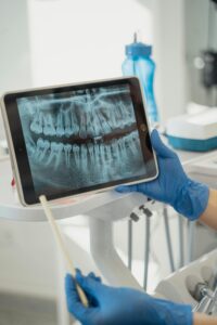
Dental Implants vs Dentures: Pros & Cons
Explore the pros and cons of dental implants vs dentures for tooth replacement in Glendale, AZ, and discover…
Read Article