Glendale, Arizona
Advanced Dental Technology in Glendale, AZ
We invest in the tools that make dentistry more accurate, more comfortable, and more predictable. From 3D cone beam imaging to digital smile design, our technology means better results -- and fewer surprises.
Why the Tools Your Dentist Uses Actually Matter
Technology is not marketing. It has direct clinical consequences. The imaging we use changes what we can see. The planning tools we use change what we can predict. The restorative equipment we use changes how long you sit in the chair and how well things fit when you leave.
Earlier Detection
Problems caught earlier are easier to treat and less expensive to fix. High-resolution imaging finds cracks, early decay, and bone changes that would be invisible on outdated equipment.
Greater Precision
Digital scans are dimensionally more accurate than physical impression trays. 3D imaging eliminates guesswork from surgical planning. Better data at the planning stage means fewer adjustments at the treatment stage.
Fewer Appointments
Same-day CAD/CAM restorations and integrated digital workflows reduce the number of visits required for many procedures. A crown that used to take two appointments now takes one.
Transparent Communication
Seeing your own X-rays and intraoral camera images on screen -- in real time -- helps you understand your oral health and participate in treatment decisions. We do not make recommendations without showing you the evidence.
Greater Comfort
Digital scanning replaces the gagging and mess of traditional impression trays. Paperless intake means no clipboard in the waiting room. A well-planned procedure is a faster, more comfortable one.
Lower Radiation
Digital radiography exposes patients to up to 70% less radiation than conventional film while producing sharper images. We take X-rays at clinically appropriate intervals, not on a fixed schedule.

Digital Dentistry
Dr. Turke's Digital-First Philosophy
Dr. Turke leads the digital dentistry program at SmileScience. His philosophy is simple: before we touch a single tooth, we should have a complete, accurate picture of the current situation and a clear, visualized plan for where we are going.
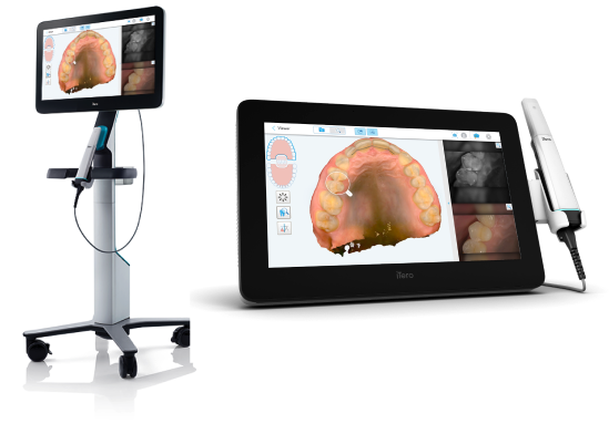
He uses the Trios 5 intraoral scanner -- one of the most accurate optical scanners available -- for digital impressions, orthodontic records, and restoration planning. Digital models replace traditional plaster impressions for most patients, meaning no impression trays, no gagging, and faster turnaround on restorations and appliances.
For cosmetic cases, Dr. Turke uses exocad and Digital Smile Design software to create a preview of proposed changes before any treatment begins. You see your potential result first -- then decide if you want to move forward.
Surgical & Implant Planning
How Dr. Dawson Uses Technology for Complex Cases
Dr. Dawson focuses on implant placement, full-arch rehabilitation, and complex oral surgical cases. For him, technology is not an option -- it is the foundation of safe, predictable treatment.
Every implant case begins with a CBCT scan that maps bone volume, density, nerve location, and sinus anatomy in three dimensions. That data is loaded into surgical planning software, where each implant is positioned digitally before a single incision is made. For full-arch cases, Dr. Dawson uses the CBCT model alongside Dr. Turke's Trios 5 scans to coordinate surgical placement with the final prosthetic design from day one.
For complex rehabilitations -- patients who need bone grafting, zygomatic implants, or full-mouth reconstruction -- the integrated digital workflow means the surgical and restorative team are working from the same data set. The result is fewer surprises, fewer complications, and a final smile that was planned before surgery began.
Technology Dr. Dawson uses for complex cases
- ✓ CBCT 3D imaging for bone evaluation and implant positioning
- ✓ Digital surgical guides from implant simulation software
- ✓ Integrated surgical-restorative planning for full-arch cases
- ✓ Nerve proximity mapping for complex extractions and grafts
- ✓ Shared data workflow with Dr. Turke for prosthetic handoff
Category 1
Imaging and Diagnostics
The images we take directly determine the accuracy of our diagnoses. We use the most capable imaging tools available in private practice dentistry.
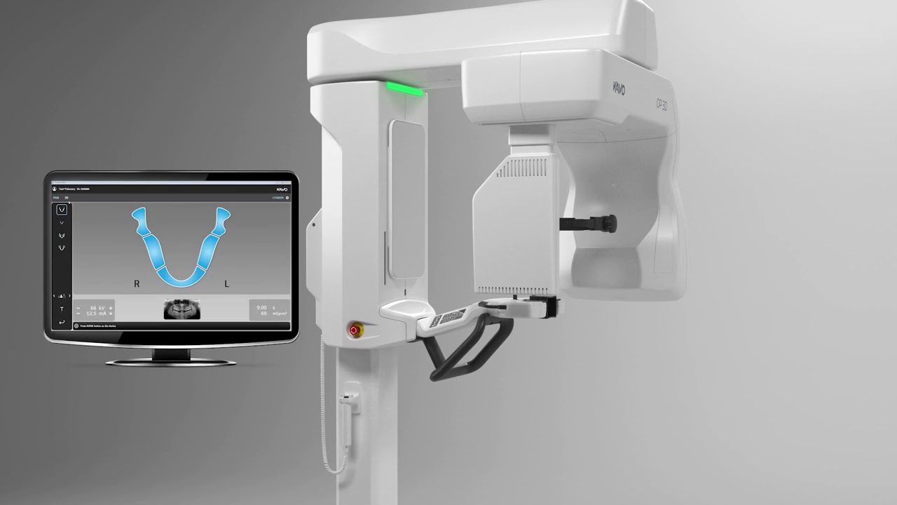
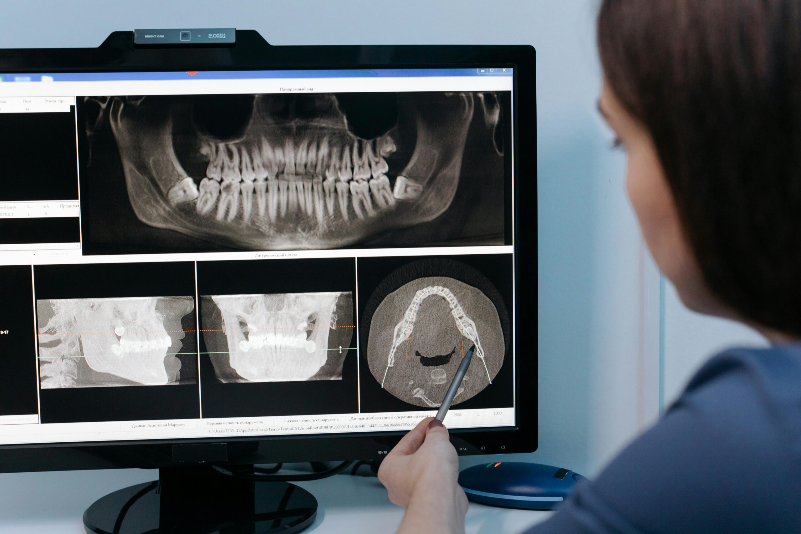
CBCT 3D Cone Beam CT Scanner
Traditional 2D X-rays show length and width. Cone beam CT adds depth -- giving us a three-dimensional view of your teeth, jawbone, nerve pathways, sinuses, and joint anatomy in a single 20-second scan. The full-arch 3D model loads on-screen instantly.
CBCT is on-site at our Glendale office. Patients do not need to visit a separate imaging center for surgical planning scans.
When we recommend a CBCT scan
- -- Implant treatment planning (bone volume, density, nerve proximity)
- -- Complex extractions (impacted teeth, root proximity to nerves)
- -- Bone graft evaluation and post-graft assessment
- -- TMJ assessment and jaw joint evaluation
- -- Root canal evaluation in complex cases
- -- Suspected pathology requiring 3D visualization
Digital X-Rays
All radiographs at SmileScience are fully digital. Digital sensors capture images in a fraction of the exposure time required by traditional film -- delivering up to 70% less radiation -- while producing sharper, higher-resolution images that load on-screen within seconds.
Images can be enlarged, adjusted for contrast, and annotated to help explain findings during your appointment. They are stored securely in your electronic record and easy to share with specialists when referrals are needed.
What digital X-rays reveal
- -- Interproximal decay (cavities between teeth)
- -- Decay beneath existing fillings or crowns
- -- Bone loss from periodontal disease
- -- Root infections, abscesses, and pathology
- -- Impacted or unerupted teeth
- -- Jaw bone density for implant planning
Intraoral Cameras
An intraoral camera is a small wand -- about the size of a pen -- that captures high-resolution video and still images of the inside of your mouth and displays them on a chairside monitor in real time.
This turns a dental examination into a shared conversation. Instead of hearing "you have a crack on tooth 14," you can see exactly what the dentist sees. Patients who understand their diagnosis are better equipped to make decisions about treatment.
What intraoral cameras help detect
- -- Cracked cusps and hairline fractures
- -- Early decay on tooth surfaces
- -- Marginal failures on existing restorations
- -- Soft tissue changes and irritation
- -- Wear patterns and occlusal damage
Category 2
Digital Treatment Planning
The "see before you commit" principle is core to how we practice. Digital planning tools let you see a proposed outcome before any irreversible treatment begins.
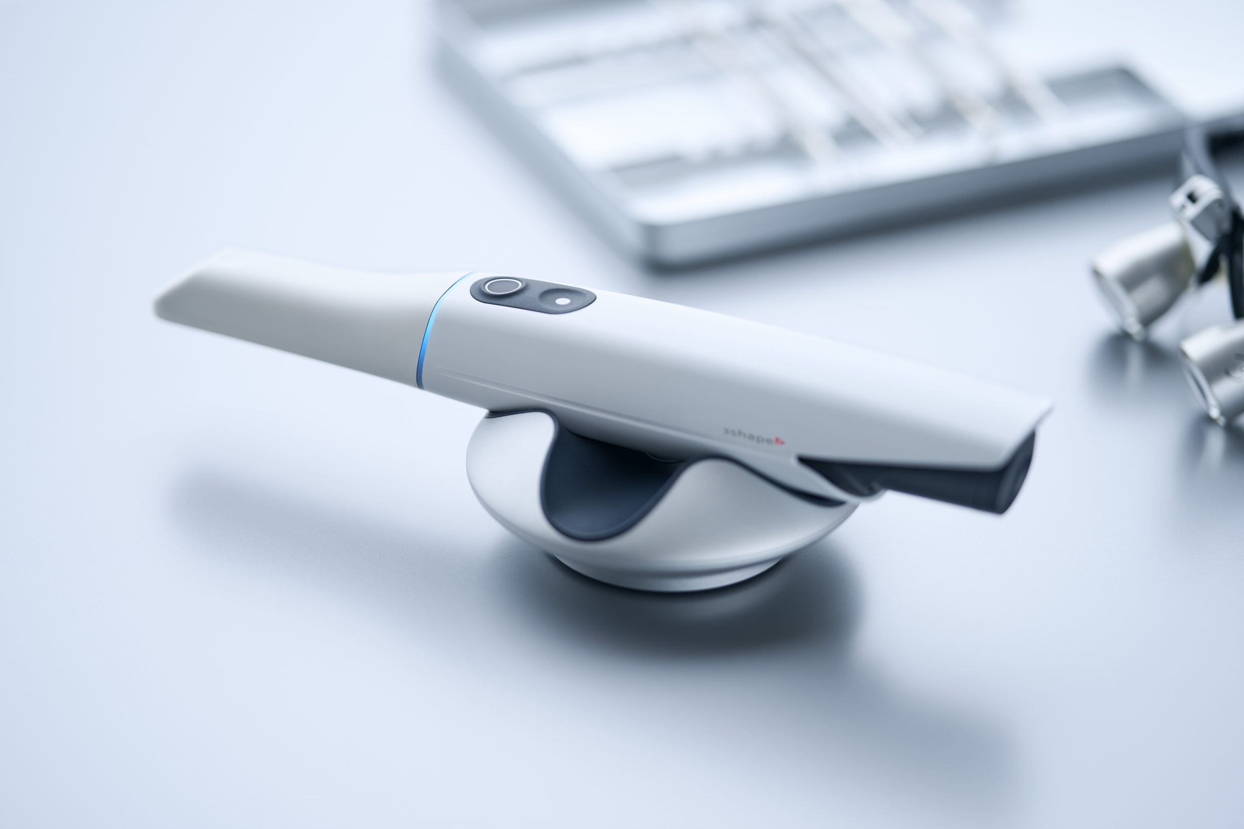
Trios 5 Intraoral Scanner
The Trios 5 is one of the most advanced optical intraoral scanners in clinical use today. Dr. Turke uses it for digital impressions, Invisalign records, veneer and crown preparation scans, and full-arch models for complex restorative cases.
No trays. No impression material. No gagging. The scanner captures a precise 3D model of your teeth in a few minutes and transmits it digitally to the lab or our in-house milling unit.
iTero Element Scanner
The iTero Element is our Invisalign-specific scanner. It captures the detailed records Align Technology requires for ClinCheck treatment simulation and aligner fabrication.
It also powers the iTero Outcome Simulator, which overlays a predicted post-treatment result on your current scan so you can see what straighter teeth would look like before committing to treatment.
ClinCheck Software
ClinCheck is Align Technology's treatment simulation platform for Invisalign. After scanning, Dr. Turke maps out each tooth movement, stage by stage, and you can watch a 3D animation of your planned tooth correction before a single aligner is made.
This means you understand the complete treatment plan -- duration, movement, and expected result -- before paying for anything.
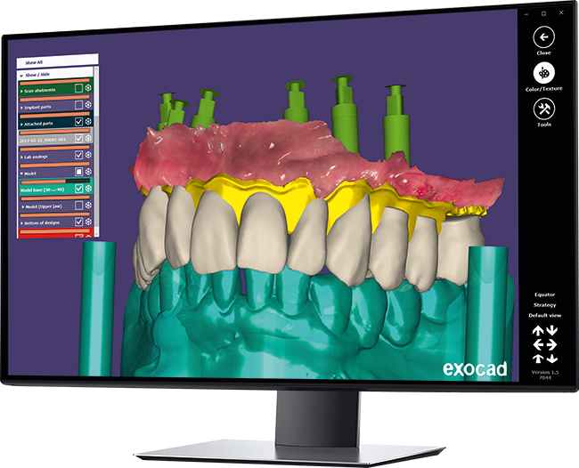
Digital Smile Design + Exocad
For cosmetic cases -- veneers, smile makeovers, full-mouth rehabilitations -- Dr. Turke uses Digital Smile Design and exocad software to create a visual preview of proposed changes before any tooth preparation begins.
This preview can be fabricated as a wax-up or temporary mockup, placed over your existing teeth, so you can literally try on your new smile before committing to permanent restorations.
Category 3
Restorative Technology
Our in-house milling and laser technology mean procedures that used to require multiple visits or outside referrals can now be completed in a single appointment at our Glendale office.
CAD/CAM Same-Day Crowns (CEREC)
Our in-house CAD/CAM milling system lets us design, mill, and place ceramic crowns, inlays, onlays, and veneers in a single appointment. No temporary crown. No second visit. No waiting two weeks for an outside lab.
The process starts with a digital scan of your prepared tooth. Design software maps the restoration to match your bite and surrounding teeth precisely. The milling unit then carves it from a solid block of dental-grade ceramic. Dr. Turke places and adjusts it the same day.
- ✓ One appointment instead of two
- ✓ No temporary that can break or fall off between visits
- ✓ All-ceramic material -- looks and feels completely natural
- ✓ Precision fit from a digital scan, not a physical impression
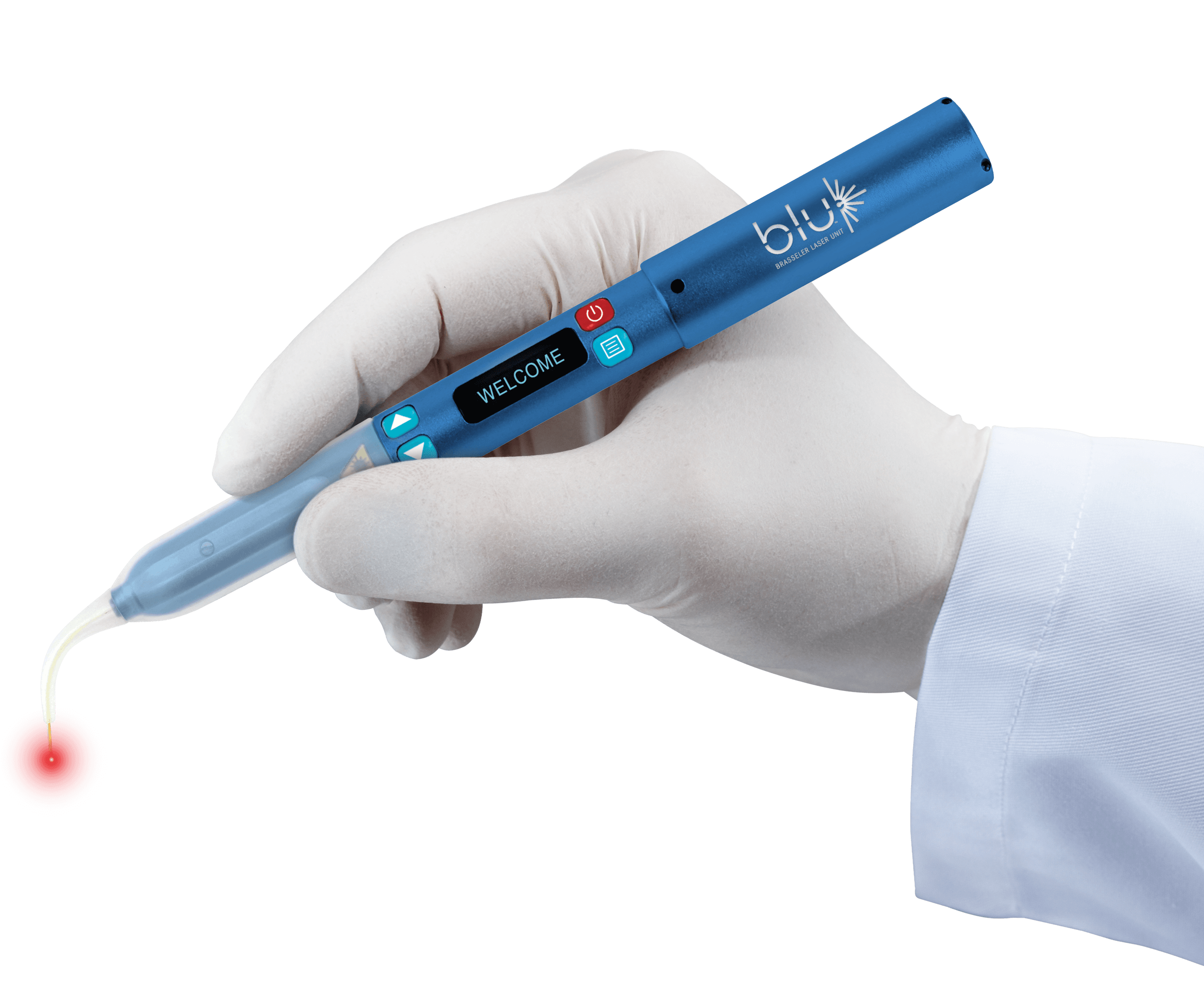
Laser Dentistry
We use soft tissue lasers for gum contouring, frenectomy, and minor periodontal procedures. Lasers seal blood vessels as they work, which means less bleeding, less post-operative swelling, and faster healing compared to traditional surgical methods.
For cosmetic cases, laser gum contouring reshapes the gumline to create a more symmetrical, proportionate smile -- often in a single visit with minimal discomfort.
- ✓ Gum contouring for cosmetic smile improvement
- ✓ Frenectomy (tongue-tie / lip-tie release)
- ✓ Soft tissue management during restorative procedures
- ✓ Faster healing vs. traditional surgical approaches
Complete Equipment List
Full Technology Inventory
Every tool in our Glendale office — what it does and who uses it.
Frequently Asked Questions
CAD/CAM ceramic crowns are milled from the same high-strength dental ceramic used by dental labs. Longevity and fracture resistance are comparable to laboratory-fabricated porcelain crowns. The key difference is convenience: one visit instead of two, and no temporary restoration that can dislodge or break between appointments.
A CBCT scan delivers a higher dose than a single periapical X-ray, but it is still a relatively low-dose procedure -- significantly below the levels used in medical CT scanning. We recommend CBCT only when the diagnostic benefit justifies the additional exposure, primarily for surgical planning, implant evaluation, and complex extraction cases. We will always explain why we are recommending one before proceeding.
Yes. We use the iTero Element scanner to capture digital records for Invisalign treatment planning. No trays, no putty, no gagging. The scan is transmitted electronically to Align Technology to generate the ClinCheck simulation, which we review with you before any aligners are ordered.
The Trios 5 is a high-precision intraoral scanner used by Dr. Turke for digital impressions for crowns, bridges, veneers, and other restorations; full-arch models for implant planning; and records for cosmetic treatment simulations. It replaces traditional impression trays entirely for most procedures and produces dimensionally more accurate models.
Yes, for cosmetic cases. Dr. Turke uses Digital Smile Design and exocad software to create a preview of proposed changes -- veneers, gum contouring, tooth reshaping -- before any tooth preparation takes place. For cases that warrant it, a physical mockup or wax-up can be created and placed in your mouth so you can literally try on the result before committing.
Soft tissue laser procedures are generally well-tolerated. Local anesthetic is used when needed. Most patients describe far less discomfort and faster healing after laser gum procedures compared to what they expected. The laser seals tissue as it works, which means minimal bleeding and swelling.
Digital X-rays produce very low levels of radiation and are generally considered safe during pregnancy when clinically necessary, especially with proper lead shielding. We always discuss your health history and pregnancy status before recommending any X-rays, and we follow ADA guidelines on appropriate use. If you are pregnant, please let us know when you schedule so we can plan your appointment accordingly.
Some do and some do not. CBCT imaging on-site, Trios 5 scanning, in-house CAD/CAM milling, and Digital Smile Design capability are not standard across all private practices. Having all of these tools in one office -- rather than referring out for imaging or lab work -- reduces your time and cost and keeps your entire care team coordinated.
Experience Modern Dentistry in Glendale, AZ
3D imaging, digital smile design, same-day restorations, and digital impressions -- all under one roof at our Glendale office. Schedule your visit to see the difference technology makes.