Pick your insurance
Sixteen carriers including Delta, Cigna, Aetna, all 4 major BCBS-network plans, AARP, and "I don't see my plan." We file claims for every carrier.
Glendale, Arizona
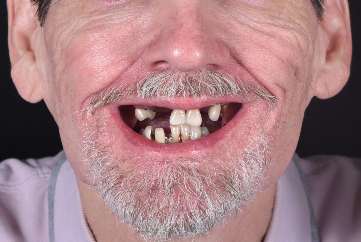
If you have been told you "don't have enough bone" for dental implants, bone grafting can rebuild the foundation your jaw needs. At SmileScience Dental Spa, Dr. Dawson evaluates your bone volume with a 3D CBCT scan and recommends the most conservative graft procedure that sets you up for a successful, lasting implant outcome.
You May Still Be a Candidate
Bone loss after tooth extraction, gum disease, or long-term denture use is extremely common -- and extremely correctable. Bone grafting rebuilds the volume and density your jaw needs to support implants successfully.
Bone Loss Begins Immediately
Within weeks of losing a tooth, the jawbone begins to shrink through resorption. Without the stimulation of a root, the body redirects minerals away from the site -- up to 25% of bone width can be lost in the first year alone.
Grafting Corrects the Deficit
Graft material placed at the deficient site acts as a scaffold. Your body's healing process replaces it with new, living bone over several months -- creating a stable site with adequate height and width for a titanium implant.
Most Patients Qualify After Grafting
Even patients with years of denture wear or significant bone loss can typically become implant candidates after appropriate grafting. A 3D CBCT scan at your free consultation gives us a precise picture of what is needed.
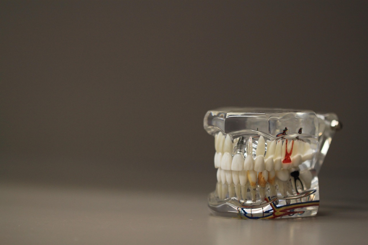
A dental implant is a titanium post that is surgically placed into the jawbone and integrates directly with living bone through a process called osseointegration. That integration requires adequate bone height, width, and density at the implant site. Too little bone means the implant has nothing to grip -- it will fail to integrate, move, or require removal.
The minimum bone dimensions for a standard implant are approximately 6 mm of width and 10 mm of height at the planned site. Many patients who had teeth removed without socket preservation years ago no longer meet these minimums without preparation. Bone grafting corrects that deficit before implant placement.
The best time to preserve bone is immediately at extraction, before resorption begins. If that window has passed, grafting can still restore volume -- the process simply takes longer. The important point: bone loss from tooth extraction is rarely permanent or irreversible.

The appropriate procedure depends on how much bone has been lost, where in the jaw the implant will be placed, and when the grafting is happening relative to tooth extraction.
Performed immediately after tooth extraction. Graft material is placed directly into the empty socket before the tissue closes over it. This is the least invasive approach and the most efficient -- it maintains bone width and height at the site while the area heals naturally.
Best for: Patients planning implants who need an extraction first.
Timeline: Implant placed 3 to 4 months after socket healing.
Used when bone resorption has already occurred and the ridge is too narrow or too short for an implant. Particulate graft material is placed against or around the deficient ridge and covered with a membrane to guide bone regeneration. Ridge augmentation restores missing horizontal and vertical dimensions.
Best for: Patients who had teeth removed months or years ago without preservation.
Timeline: 4 to 6 months before implant placement.
Indicated for larger horizontal defects where particulate graft material alone is not sufficient. A block of bone -- most commonly from the patient's own jaw or a processed donor source -- is secured to the deficient area with small titanium fixation screws. Block grafts are the most aggressive approach and reserved for significant deficits.
Best for: Significant horizontal bone deficiency requiring large-volume augmentation.
Timeline: 4 to 6 months of integration before implant placement.
A specialized graft for the upper back jaw, where the sinus cavity sits close to the ridge. The sinus membrane is gently elevated and bone graft material is placed beneath it to build height for implants in the upper molar region. This is the most common grafting procedure needed for upper jaw implants.
Best for: Missing upper molars or premolars with insufficient vertical bone height.
Timeline: 4 to 9 months depending on approach and graft volume. Learn more about sinus lifts.
Platelet-Rich Fibrin (PRF) is a concentration of growth factors drawn from your own blood at the time of surgery. Added to or layered over graft material, PRF accelerates healing, reduces inflammation, and improves graft integration outcomes. SmileScience uses PRF routinely for grafting procedures.
Best for: Any graft case where faster integration or reduced post-op swelling is a priority.
Timeline: Integrated into the graft appointment -- no additional visit required.
In select cases where existing bone height is adequate in one dimension but slightly deficient in another, grafting and implant placement can be performed simultaneously. This approach reduces total treatment time when appropriate bone contact can still be achieved for initial implant stability.
Best for: Patients with minor ridge deficiency adjacent to adequate bone.
Timeline: Single surgical appointment; healing mirrors standard implant timeline.
Several sources of bone graft material are clinically proven and routinely used. The best choice depends on the size of the defect, the patient's anatomy, and the surgical approach.
Autograft -- Your Own Bone
Bone harvested from another site in the patient's own body -- commonly from the jaw itself (chin or ramus), hip, or tibia. Autograft is the biological "gold standard" because it contains living cells that integrate readily. The tradeoff is a second surgical site and a slightly longer recovery. Used most often for large block grafts requiring significant volume.
Allograft -- Processed Human Donor Bone
The most common choice for socket preservation and ridge augmentation. Allograft comes from a tissue bank, is processed to remove all cellular material (making it safe), and provides an excellent scaffold for new bone growth. No second surgical site is required. Long-term studies show high success rates comparable to autograft for most applications.
Xenograft -- Animal-Derived Bone
Most commonly bovine (cow) or porcine (pig) derived. Like allograft, it is extensively processed and sterile. Bovine xenograft (such as Bio-Oss) is widely studied and resists resorption very well, making it a strong choice for sinus lifts and sites where long-term volume maintenance is important. No additional surgery is required.
Alloplast -- Synthetic Bone Substitute
Synthetic materials such as hydroxyapatite, tricalcium phosphate, or bioactive glass. These materials have no biological source and are a good option for patients who prefer to avoid donor or animal materials for personal or religious reasons. Modern synthetics perform well in many applications, particularly when combined with PRF.
A cone-beam CT scan gives Dr. Dawson a three-dimensional view of your bone volume and the precise location of anatomical structures like the sinus cavity and inferior alveolar nerve. The scan also reveals the quality of remaining bone and guides both graft design and the eventual implant placement plan. This imaging is included free at your consultation.
The surgical site is numbed thoroughly with local anesthesia before anything begins. For patients who prefer a more relaxed experience, oral conscious sedation or IV sedation administered by a board-certified dental anesthesiologist is available. You will not feel pain during the procedure regardless of which approach is chosen.
A small incision is made in the gum tissue to access the underlying bone. The site is cleaned and shaped to receive the graft material. For autograft block procedures, the donor site is prepared separately.
Graft material is carefully packed into the deficient site. In most cases, a collagen or resorbable membrane is placed over the graft. PRF may be layered in to accelerate healing. The tissue is sutured closed.
Over the next 3 to 9 months, your body gradually replaces the graft scaffold with your own living bone. A follow-up CBCT scan at the appropriate interval confirms bone density and readiness before implant surgery is scheduled.
The total time from graft to implant placement ranges from 3 to 9 months depending on the type of graft and your body's healing rate.
Peak swelling and soreness. Rest, soft diet, cold compresses, and prescribed medications manage discomfort. Most patients find this period comparable to a tooth extraction.
Swelling resolves. Sutures dissolve or are removed at a follow-up visit. Return to most normal activities. Soft diet continues until Dr. Dawson clears full function.
Socket preservation and smaller ridge augmentation sites are typically ready for implant evaluation. A follow-up CBCT scan confirms bone density.
Block grafts, large ridge augmentations, and sinus lifts require additional integration time. The wait is necessary for long-term implant stability.
While the healing window can feel long, patients consistently report that the wait is worth it. Implants placed in well-grafted bone have the same long-term success rates as implants placed in native bone -- the extra time simply ensures the same quality foundation.
Grafting costs vary depending on the procedure type, graft material, and the number of sites being treated.
Typically ranges from $300 to $800 per site when performed at the time of extraction. This is the most cost-effective grafting approach because it prevents a more involved (and more expensive) ridge augmentation procedure later.
Ranges from $600 to $3,000 depending on defect size, graft material, membrane type, and whether PRF is used. Multiple adjacent sites may qualify for combined fee structures.
Transcrestal (minimally invasive) sinus lifts range from $700 to $1,500 per side. Lateral window sinus lifts range from $1,500 to $3,000 per side. Both are significantly more affordable than the cost of not placing implants and dealing with long-term denture complications.
Many dental insurance plans include some coverage for bone grafting, particularly socket preservation following extraction. FSA and HSA accounts cover grafting costs in full. We also offer financing through CareCredit, Cherry, Sunbit, and Proceed Finance, with monthly options available. Ask our team about your specific options at your consultation.
Live coverage estimator
Three quick taps — pick your insurance, pick your procedure, see the patient-cost range against our 2026 fee schedule next to our in-house membership plan. No login, no email, no sales pitch.
Estimate range, not a quote. Final cost depends on your specific plan benefits, remaining annual max, and clinical findings at your consult. Defaults used here: $1,500 annual max, $50 deductible.
Sixteen carriers including Delta, Cigna, Aetna, all 4 major BCBS-network plans, AARP, and "I don't see my plan." We file claims for every carrier.
Plain-language categories — Cleaning, Filling, Crown, Root Canal, Extraction, Gum Treatment, Implants, Cosmetic. Drill down to the specific option that matches your case.
Insurance estimate vs. our in-house membership plan, side-by-side. Ranges (not single numbers) so you're not surprised at the desk. Real 2026 Glendale-market pricing.
No. Patients who have sufficient bone height, width, and density can proceed directly to implant placement without grafting. A cone-beam CT scan is the definitive way to determine whether grafting is needed. Many patients who lost a tooth recently and had socket preservation performed at the time of extraction have adequate bone for a straightforward implant placement.
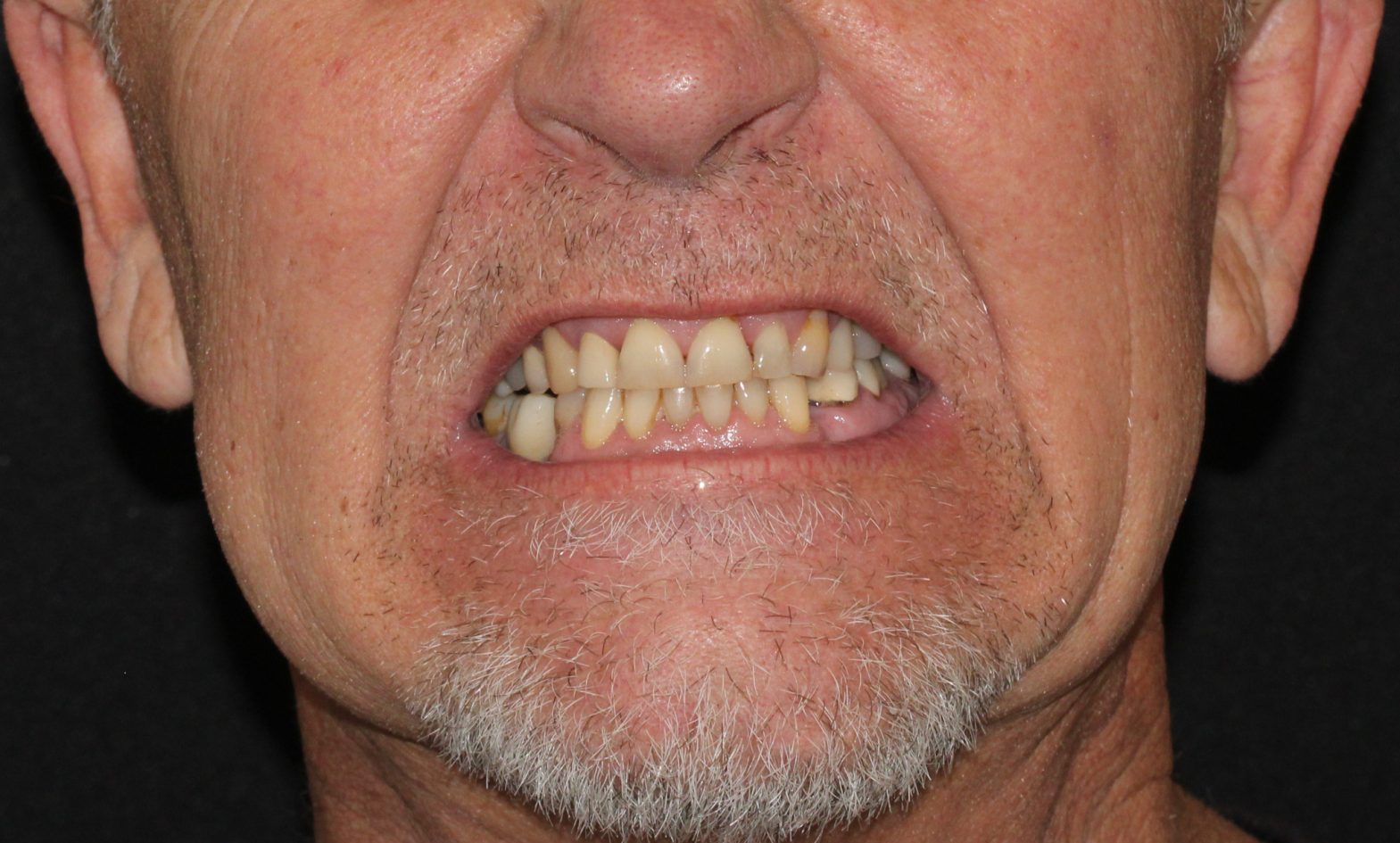
Usually yes -- but with appropriate preparation. Long-standing bone loss simply means more grafting is required before implants can be placed. Even patients who have worn full dentures for 10 or 20 years and have significant bone atrophy often remain candidates for implant-supported restorations once adequate bone volume is restored. The evaluation starts with a 3D CBCT scan to see exactly what is there.
Several options exist and the choice depends on your specific case. Allograft (processed human donor bone) is the most common, with an excellent track record and no additional surgical site required. Xenograft (bovine or porcine-derived) is widely used for sinus lifts and ridge augmentations where long-term volume is important. In cases requiring a block graft, autograft (the patient's own bone) may be harvested from the jaw. Synthetic alloplasts are available for patients who prefer to avoid biological materials. Dr. Dawson will explain the rationale for your specific situation.
Most patients describe the first 2 to 3 days as comparable to a tooth extraction -- soreness and mild to moderate swelling that is well managed with over-the-counter anti-inflammatories and, when appropriate, a short course of prescription medication. Patients who receive IV sedation typically report minimal post-operative discomfort and are surprised by how comfortable the experience was. By day 4 to 5, most patients are back to a near-normal routine.
Graft failure -- where the graft does not integrate -- is uncommon with proper surgical technique and patient compliance, but it can occur. The most common causes are smoking, infection, and failure to follow post-operative instructions (particularly regarding diet and oral hygiene). If a graft does not integrate successfully, the site can typically be re-grafted after it heals. The outcome is not permanent -- it is a setback that can be addressed.
Yes, significantly. Smoking reduces blood flow to healing tissue and impairs the body's ability to form new bone. Smokers have substantially higher rates of graft failure and implant failure than non-smokers. Dr. Dawson will discuss this candidly during your consultation. We do not refuse care to smokers, but we do require honest informed consent about the impact on outcomes and typically recommend smoking cessation programs as part of a successful treatment plan.
Coverage varies widely. Some dental plans cover socket preservation as part of an extraction benefit, particularly when it is performed at the time of tooth removal. Standalone ridge augmentation is less commonly covered. Our team will verify your benefits before treatment and provide a clear breakdown of estimated out-of-pocket costs. FSA and HSA accounts cover grafting in full, and we offer financing for the remaining balance.
The graft material begins to integrate with your existing bone within the first few weeks, but mature bone density takes longer to develop. Socket preservation sites are typically ready for implant placement in 3 to 4 months. Ridge augmentations require 4 to 6 months. Sinus lifts typically need 4 to 9 months depending on the volume grafted. A follow-up CBCT scan confirms density before implant surgery is scheduled -- we do not operate on timelines alone.
Sometimes. In cases where existing bone provides enough height and density for initial implant stability -- even if supplemental grafting is needed around it -- simultaneous placement is possible and reduces overall treatment time. This is evaluated case by case during the CBCT review. When in doubt, Dr. Dawson errs toward staged placement to maximize long-term integration success.
4.9 (437 reviews)
I recently had the pleasure of visiting Smile Science Dental Spa, and I couldn't be more impressed with the level of care and service provided by the entire team. Dr. Dawson and Dr. Turke are both incredibly skilled and made me feel completely at ease throughout my procedures. Their professionalism, combined with their gentle approach, made me feel comfortable and well-informed every step of the way. I was especially impressed that I was able to get a same-day crown(apparently their first ever!), which exceeded my expectations. I couldn’t believe how quick and seamless the process was. It really saved me time and made the experience even more convenient. Cindiray and Angeles, the dental assistants, were equally wonderful. They were not only friendly and attentive but also thorough in explaining everything and made sure I was relaxed during each phase of my visit. Lastly, a big thank you to Litzy at the front desk, who greeted me with a smile and helped with scheduling and any questions I had. She made the entire check-in process a breeze. From start to finish, my experience at Smile Science Dental Spa was exceptional. I'll be returning for sure. I highly, highly, highly recommend them for anyone looking for high-quality dental care.
View Full Review on GoogleDusty
Honestly I’ve never been so happy to go to the dentist. I came in (late due to Lyft) and was still taken care of and treated with care. Even without insurance (kicking in within the next month) prices for an extraction on my infected tooth were more than reasonable. I was able to get more than expected done in the same visit. Dr. Dawson and his team made me feel so comfortable and taken care of. Won’t go anywhere else.
View Full Review on GoogleCherish B
I recently started as a patient after leaving our long-time family Dentist and couldn’t be happier! Marcela is AMAZING and a FANTASTIC hygienist!!!! 😃 Dr Dawson is great too! Every time I visit, they make me feel like family.
View Full Review on GoogleTJ Winzeler
Myself and my family had previously gone to Dr. Turk at another dental office that he worked at. He had relocated and we did not know where he had gone to until this week. Today I got in with Dr. Turk and he took the time to look at my x-rays carefully . I was having pain from a tooth and some other issues. The previous Dentist I went to that I saw just a few days earlier, he did not find any issues with my tooth which I had pain in. Dr. Turk identified infection and referred me to an endodontist. He is very gentle as a wonderful disposition and my self and my family are thrilled to be able to get back with him. The office is lovely, relaxing music, heat and massage chair.I have always been very nervous and some anxiety going to dentist as I have had bad reactions with the anesthesia. I am confident that Dr. Kirk will help me get through my next appointment just fine. “The staff are very kind and attentive. I am looking forward as I have quite a bit of work to be done, and I know I’m in good hands with Dr. Turk and his staff. Five stars!⭐️ Sherry
View Full Review on GoogleSherry Redmond
I recently moved to Arizona and had an issue with a tooth repair from 15 years back. Not knowing anyone here in my new home, I searched dentists close to me. I chose Smile Science much like pinning the tail on the donkey and my selection couldn't have been better! What a terrific staff! Dr. Turke was concerned the tooth would need to be extracted, but said he wanted to take a shot at saving it..and that's just what he did! After setting a temporary crown, I was on my way with an appointment to install the permanent crown in the following two weeks. That evening I received a call I've never received from a dentist. Dr. Turke had taken the time to follow up with me to make sure I wasn't in any discomfort..I was completely caught off guard by this kind of attention to detail and caring for my well being. The permanent crown is now in place, it fits and feels fantastic, the job and treatment are first class! Thank you, Dr. Turke and staff for taking in this stray and giving me a home dentist! I'd like to say that I'm just good at selecting blindly, but the truth is I am very lucky..and I'll take lucky over being good any day of the week! Thank you, again and Happiest of New Year's in 2026, Science Smile!!
View Full Review on GoogleEddy
Schedule a free bone grafting consultation at SmileScience Dental Spa in Glendale, AZ. Dr. Dawson will review your CBCT scan, confirm whether grafting is needed, explain exactly what procedure applies to your case, and map out a timeline from graft to final restoration.


